
Understanding Tendon Pain
Part I - Tendon Structure and causes of pain
To understand what tendon pain is and how it’s treated, it’s important to understand how it begins.
What are tendons?
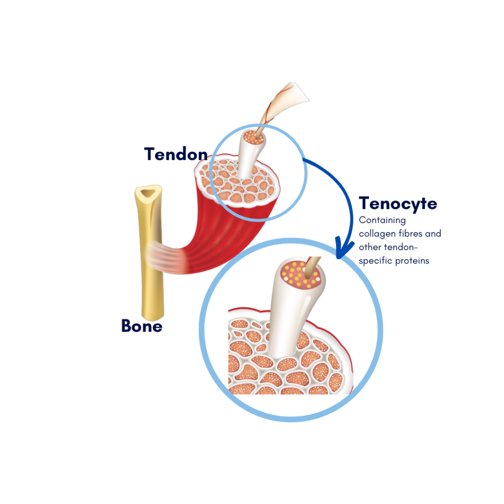
Tendons are structures in the body which connect muscle to bone, helping your bones and muscles to move together and bear loads safely.
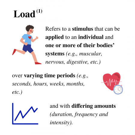
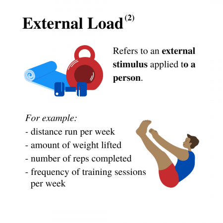
Important terms for understanding the role of tendons:
Healthy tendons are comprised of:
- Tendon-specific cells (tenocytes)
- Collagen (Type I)
- Proteins (proteoglycans)
- Other complementary elements (3)
Healthy tendons keep their structure by balancing tissue breakdown and production. Under or overloading your tendons can lead to changes in their structure (4). This can cause pain on, or around the tendon.
Underloading refers to rest longer than 2-3 weeks leads where the tendon is being used less than normal.
This can lead to a reduction in the tendon’s capacity, potentially causing it to overload (5).
Overload is defined as when the tendon’s capacity to bear load is less than the load is it being put under. In other words, it can’t meet the needs of the activities you’re doing. This occurs through either the tendon resting and starting to breakdown, or an increase in activity too fast for what the tendon can adapt to.
This is relevant for people who have been wearing a brace, had surgery, or have simply stopped exercising as frequently during winter. Those who dramatically increase their activity levels after a rest period are at risk of tendon breakdown, which occurs when the activity continues to exceed the capacity of the tendon.
Acute overload causes changes and loosening in tendon structure (also known as the tendon matrix). This leads to the degeneration of the tendon and as a result, type 1 collagen in the tendon is replaced with type II and III, which is not as effective at bearing load.
This stage of tendon injury is most likely irreversible; however the tendon can function well and pain-free with an effective exercise program and education. When activity is reduced back to what the tendon can handle, the tendon responds by increasing the amount of normal tendon tissue around the area. This maintains its ability to meet the demands of the activity.
Key Take Homes:
- Load changes are always involved with tendon injury. Both under and/or overloading can lead to changes in tendon structure.
- As recovery from tendon injury continues, the tendon adapts to irreversible change by adding more normal tendon tissue. This means the tendon can tolerate the demands of rehabilitation and with guidance, you can safely return to your activity symptom-free.
If you’ve been diagnosed with a tendon injury or think you may be suffering from tendon pain, make an appointment with one of our experienced physiotherapists and get back to exercising pain-free!
Stay tuned for Part Two – Assessment & Diagnosis of Tendon Injuries
Zac explains how City Baths Physio practitioners assess and diagnose tendon pain, with some tips on symptoms of tendon pain to watch out for.
Stay in touch & up to date via our socials!
References:
1. Soligard T, Schwellnus M,
2. Halson SL. Monitoring training load to understand fatigue in athletes. Sports Med 2014;44(Suppl 2):S139–47
3. Sprague AL, Smith AH, Knox P, Pohlig RT, Sibernagel KG. Modifiable risk factors for patellar tendinopathy in athletes: a systematic review and meta analysis. British Journal of Sports Medicine. 2018;52(24): 1575-85
4. Rosso F, Bonaisa DE, Cottino U, Dettoni F, Bruzzone M, Rossi R. Patellar tendon: From Tendinopathy to rupture. Asia-Pacific Journal of Sports Medicine, Arthroscopy, Rehabilitation and Technology. 2015;2(4): 99-107
5. McAuliffe S, Tabuena A, McCreesh K, O’Keeffe M, Hurley J, Comyns T, et al. Altered strength profile in Achilles Tendinopathy: A systematic review and meta-analysis. Journal of Athletic Training. 2019;54(8): 889-900